
Contact us: tneogi@bu.edu
Contact us: tneogi@bu.edu
|
This web-based calculator is intended to facilitate the scoring of the ACR- and EULAR-endorsed “ACR-EULAR Gout Classification Criteria”, published in Arthritis & Rheumatology1 and Annals of the Rheumatic Diseases2 in 2015. These criteria are intended for identifying subjects who may be eligible for entry into a clinical study; they should not be used for diagnosis.3 Full details regarding how to use this instrument can be found in the publications.1,2 In brief, these criteria should only be scored if the subject meets the entry criterion. If the subject meets the sufficient criterion, the subject may be classified as having gout without further scoring. For each domain, the items are listed in hierarchical order and are mutually exclusive. As such, each domain should be scored for the highest category ever noted for the subject. A score of ≥8 classifies a subject as having gout. The full ACR-EULAR Gout Classification Criteria table with values for each item’s score can be downloaded here. Information entered into this web page will not be stored. You may print the results for your records. References:
| |||
Entry CriterionThese criteria only apply to those who meet the Entry Criterion |
At least one episode of swelling, pain, or tenderness in a peripheral joint or bursa | ||
| Subject is not eligible for scoring | |||
Sufficient Criterion(If met, can classify as gout without applying criteria below) |
Presence of MSU crystals in a symptomatic joint or bursa (i.e., in synovial fluid) or tophus | ||
| Subject meets sufficient criterion and can be classified as having gout | |||
| Thank you. Assessment of this subject’s classification for gout is now complete | |||
Criteria (to be used if Sufficient Criterion not met):Score ≥8 required for classification as gout |
Categories Please select the highest category ever noted for each criteria. |
Select | |
| Pattern of joint/bursa involvement during symptomatic episode(s) ever | Joint(s) or bursa(e) other than ankle, midfoot or 1st MTP (or their involvement only as part of a polyarticular presentation) | ||
| Ankle OR midfoot (as part of monoarticular or oligoarticular episode without MTP1 involvement) | |||
| MTP1 (as part of monoarticular or oligoarticular episode) | |||
Characteristics of symptomatic episode(s) ever:
|
No characteristics | ||
| One characteristic | |||
| Two characteristics | |||
| Three characteristics | |||
| Time-course of episode(s) ever:
Presence (ever) of ≥2, irrespective of anti-inflammatory treatment:
|
No typical episodes | ||
| One typical episode | |||
| Recurrent typical episodes | |||
Clinical evidence of tophus:

Clinical evidence of tophus. Examples of tophus at typical sites: A. Ear, B. elbow (olecranon bursa), C. and D. finger pulps. Draining or chalk-like subcutaneous nodule under transparent skin, often with overlying vascularity, located in typical locations: joints, ears, olecranon bursae, finger pads, tendons (e.g., Achilles). |
Absent | ||
| Present | |||
| Serum urate: Measured by uricase method. Ideally should be scored at a time when the patient was not taking urate-lowering treatment and patient was beyond 4 weeks of the start of an episode (i.e., during intercritical period); if practicable, retest under those conditions. The highest value irrespective of timing should be scored. | <4mg/dL [<0.24mM] | ||
| 4-<6mg/dL [0.24-<0.36mM] | |||
| 6-<8mg/dL [0.36-<0.48mM] | |||
| 8-<10mg/dL [0.48-<0.60mM] | |||
| ≥10mg/dL [≥0.60mM] | |||
| Synovial fluid analysis of a symptomatic (ever) joint or bursa: Should be assessed by a trained observer. | Not done | ||
| MSU negative | |||
|
Imaging evidence of urate deposition in symptomatic (ever) joint or bursa: Ultrasound evidence of double-contour sign 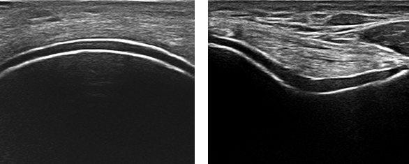
Ultrasound double contour sign. Left panel shows longitudinal ultrasound image of the femoral articular cartilage and right panel shows transverse ultrasound image of the femoral articular cartilage. Both images show hyperechoic enhancement over the surface of the hyaline cartilage. We gratefully acknowledge Dr. Esperanza Naredo, Hospital Universitario Gregorio Maranon, Madrid, Spain for providing these images. DECT demonstrating urate deposition. 
Dual energy computed tomography (DECT) urate deposition. Left panel shows urate deposition at the 1st and 5th metatarsophalangeal joints, and right panel show urate deposition within the Achilles tendon. |
Absent OR Not done | ||
| Present (either modality) | |||
Imaging evidence of gout-related joint damage:
Conventional radiography of the hands and/or feet demonstrate at least one erosion.

Imaging evidence of gout-related joint damage. Bone erosion on plain radiography in the 1st metatarsophalangeal joint, defined as a cortical break with sclerotic margin and overhanging edge. |
Absent OR Not done | ||
| Present | |||
| TOTAL SCORE | 0 | ||
| CLASSIFY AS GOUT? | |||